How Dentists Diagnose and Plan Gum Disease Treatment
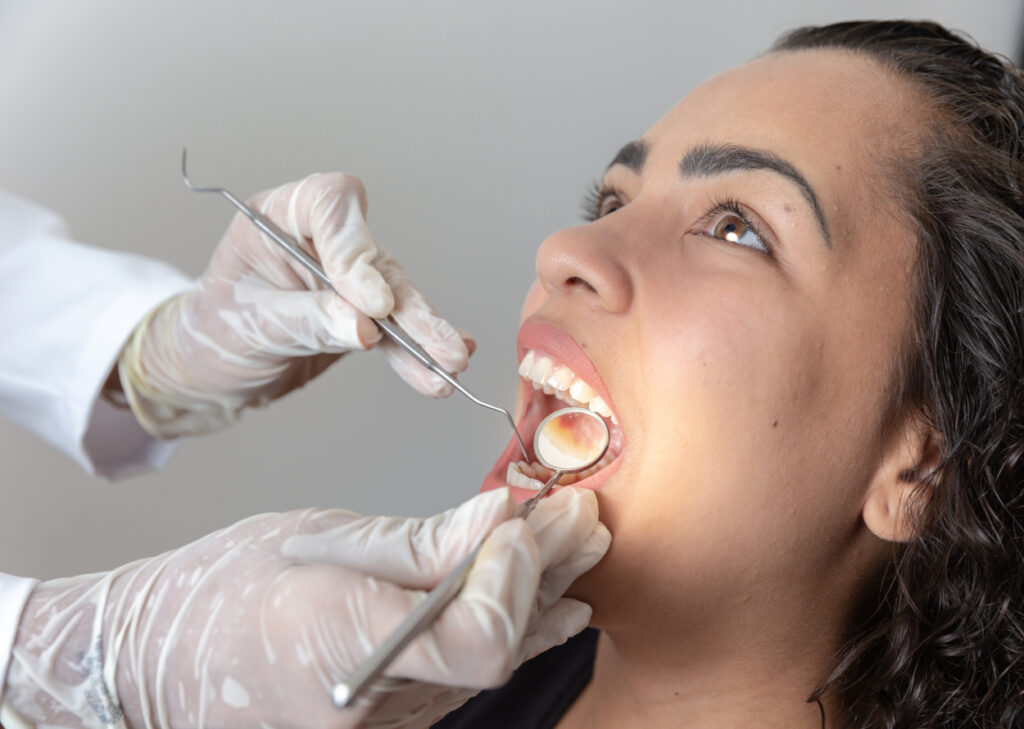

Gum disease rarely announces itself with drama at the start. More often, it begins with small signs patients live with for months, sometimes years. A little bleeding when flossing. Breath that never quite feels fresh. Tender gums around one back tooth. Spacing that seems slightly different than it used to be. By the time discomfort becomes obvious, the infection may already be affecting the supporting structures that keep teeth stable.
That is why diagnosis matters as much as treatment. Effective Gum Disease Treatment is not just a cleaning with stronger tools. It starts with careful observation, good records, and a realistic understanding of how the disease behaves in a specific mouth. Two people can both be told they have periodontal disease and still need very different plans. One may respond well to non-surgical therapy and tighter home care. Another may need staged treatment, bite adjustment, grafting, or referral to a periodontist. The difference lies in the details dentists gather before the first instrument ever touches the gums.
Why gum disease is more complex than it looks
Patients often think of gum disease as an all-or-nothing problem. Either the gums are healthy or they are infected. In practice, it exists on a spectrum. Gingivitis is inflammation confined to the gum tissue. Periodontitis means the supporting attachment around the tooth has been damaged. Once attachment and bone are lost, the body does not simply rebuild those structures on its own.
The challenge is that symptoms do not always match severity. I have seen patients with dramatic redness and swelling but little permanent damage, and others with almost no pain who had deep pockets and silent bone loss. Smokers, in particular, can be deceptive. Nicotine constricts blood vessels, so gums may bleed less even while disease progresses underneath. Patients with diabetes can show another pattern, where inflammation is more aggressive and healing less predictable if blood sugar is poorly controlled.
This is one reason experienced dentists do not diagnose gum disease by quick visual impression alone. They look at patterns, compare findings tooth by tooth, and ask whether the tissue changes make sense for the patient’s age, health history, habits, and past dental work.
The first clues usually come from the exam room conversation
A thorough diagnosis starts before the periodontal probe comes out. The medical and dental history often tells the dentist where to look more closely. Some of the most useful information comes from simple questions asked well.
Bleeding is an obvious clue, but not all bleeding means advanced disease. Someone who has not flossed in months may have inflamed gums that improve quickly with proper cleaning. On the other hand, a patient who says, “My gums bleed every time, no matter what I do,” raises a different concern. So does someone who reports loose teeth, drifting front teeth, gum tenderness in one area, a bad taste, or recurrent swelling.
Systemic health matters. Diabetes, immune disorders, pregnancy-related hormonal changes, dry mouth from medications, and smoking all influence periodontal health. A history of clenching and grinding matters too, because excessive bite forces can worsen mobility and complicate how bone loss presents on x-rays. Even family history can be useful. Some people are simply more susceptible to periodontal breakdown despite reasonable home care.
Patients are often surprised that stress, inconsistent recalls, and old dental restorations enter the conversation. They should. A crown with a rough margin or a filling that traps plaque can turn one area into a chronic trouble spot. A stressful period in life can lead to skipped hygiene visits and poor home care, and those missed months add up.
What dentists look for during the clinical exam
The visual exam provides the first direct evidence of disease activity. Healthy gums are usually firm, pale to coral pink depending on natural pigmentation, and closely adapted around the teeth. Diseased gums may appear swollen, shiny, redder than usual, or puffy at the margins. Those are classic signs, but they are only the beginning.
Dentists also assess the shape and contour of the tissue. Inflamed gums often lose their crisp architecture. The little scalloped edges between teeth flatten out. In more advanced cases, recession exposes root surfaces and creates a longer-looking tooth. That recession can be caused by periodontal disease, brushing trauma, thin tissue type, or a combination of factors, so it has to be interpreted carefully.
Plaque and tartar deposits matter because they help explain the environment that allows disease to persist. Plaque is the sticky bacterial film. Tartar, also called calculus, is hardened plaque that cannot be removed with a toothbrush. Calculus is especially important because it creates a rough surface that shelters more bacteria below the gumline.
Mobility is another finding that shapes treatment planning. A slightly mobile tooth in a patient with generalized inflammation may tighten after treatment. A mobile tooth with severe bone loss, a crack, or traumatic bite forces has a more guarded outlook. Dentists do not judge that with guesswork. They compare movement, location, and radiographic support before discussing prognosis.
Then there is furcation involvement, one of the more technical but important parts of diagnosis. Back teeth with multiple roots can lose bone between those roots. When that happens, cleaning becomes harder for both patient and clinician, and long-term maintenance becomes more demanding. Furcations often change the conversation from a simple cleaning problem to a structural one.
The periodontal probing exam, where the map gets drawn
Periodontal probing is the core measurement system for diagnosing gum disease. A slender, marked instrument is gently slid between the tooth and gum to measure the depth of the sulcus or pocket. In a healthy mouth, those measurements are often around 1 to 3 millimeters. Deeper readings can suggest loss of attachment, swelling, or both.
This part of the appointment can feel tedious to patients because the dentist or hygienist records six measurements around each tooth. There is a reason for that level of detail. Gum disease is rarely symmetrical. One tooth may have normal readings on the cheek side and a 6 millimeter pocket on the tongue side. A generalized statement like “your gums are a little inflamed” misses what actually needs treatment.
Bleeding on probing is just as significant as the numbers. A 4 millimeter site that bleeds repeatedly tells a different story than a stable 4 millimeter site with no bleeding and good home care. Suppuration, meaning pus from a pocket, is more concerning still and suggests active infection. Dentists also record recession, which allows them to calculate clinical attachment loss, a more meaningful indicator of past damage than pocket depth alone.
A few key measurements guide the next step:
- Pocket depth, which reflects how deep the space is around the tooth.
- Bleeding on probing, which signals ongoing inflammation.
- Recession, which shows how much root surface is exposed.
- Clinical attachment loss, which helps determine the true extent of support lost.
- Furcation and mobility findings, which affect prognosis and treatment complexity.
These measurements form a periodontal chart, essentially a map of the disease. That map is what allows treatment to be tailored rather than improvised.
X-rays reveal what the gums cannot show on the surface
If probing is the map, radiographs are the structural blueprint. Dentists use x-rays to assess bone levels around teeth, identify patterns of bone loss, and rule out other problems that can mimic or worsen periodontal disease.
Horizontal bone loss, where the bone height drops fairly evenly, suggests a different pattern than vertical defects, where deeper angular loss forms beside specific teeth. Vertical defects can sometimes respond well to regenerative procedures if the anatomy is favorable. Horizontal loss often calls for a more maintenance-focused strategy. That distinction cannot be made from the surface alone.
X-rays also help detect retained tartar below the gumline, open contacts that trap food, overhanging restorations, root anatomy challenges, abscesses, and cracks. In a patient with a loose tooth, the x-ray may show whether the problem is mostly periodontal, mostly endodontic, or a combination. That matters because a tooth that seems “gum related” might actually need root canal treatment, extraction, or both.
There are limitations, and good dentists remember them. X-rays are two-dimensional images of three-dimensional structures. Early soft tissue inflammation will not show. Mild bone changes can be subtle. That is why radiographs support the diagnosis, they do not replace the clinical exam.
Staging and grading, the framework behind modern diagnosis
Periodontal diagnosis has become more structured in recent years. Many dentists now classify disease using staging and grading. Patients do not need to memorize the terminology, but the concepts are useful because they explain why one treatment plan sounds more involved than another.
Staging describes severity and complexity. It looks at factors such as attachment loss, bone loss, probing depths, furcation involvement, and whether teeth have already been lost due to periodontal disease. A mild case may be limited to early tissue support loss. A severe case may include deep pockets, drifting teeth, bite collapse, and reduced chewing function.
Grading estimates how fast the disease is progressing and what risk factors are pushing it. Smoking and uncontrolled diabetes can shift a case into a higher-risk category even if the current damage looks moderate. That affects prognosis, recall intervals, and how firmly the dentist emphasizes risk reduction before or alongside treatment.
This framework helps prevent under-treatment and over-treatment. Without it, a patient with isolated 4 millimeter pockets might be managed the same way as someone with generalized 7 millimeter pockets, furcation defects, and active smoking. Clinically, those are very different situations.
Not every bleeding gum needs the same kind of care
One of the most important parts of diagnosis is deciding what the inflammation actually represents. Patients often hear terms like “deep cleaning” used broadly, but a responsible dentist distinguishes between routine preventive cleaning, gingivitis therapy, and treatment for periodontitis.
A routine cleaning is for a generally healthy mouth, where plaque, light calculus, and superficial staining are removed above and slightly below the gumline. It is preventive care, not disease management.
Gingivitis therapy may involve more focused cleaning and coaching because the tissue is inflamed, but the attachment and bone support have not been permanently lost. Many of these cases improve dramatically in a few weeks if plaque control becomes consistent.
Periodontitis requires a different level of intervention because the infection has already affected the supporting structures. That usually means scaling and root planing, often done by quadrant and sometimes with local anesthetic, followed by reassessment. If deeper pockets remain, surgery or specialist care may be needed.
There is a practical reason dentists separate these categories. If the diagnosis is wrong, the treatment either falls short or becomes unnecessarily aggressive.
How dentists build a treatment plan that fits the patient
Once the diagnosis is clear, the treatment plan takes shape. This is where technical findings meet real life. A perfect plan on paper is useless if the patient cannot tolerate it, afford it, maintain it, or return for follow-up.
Dentists weigh several things at https://jaredrind922.theburnward.com/the-recovery-timeline-after-gum-disease-treatment once. Severity is the obvious one, but not the only one. They also consider the distribution of disease. Generalized moderate periodontitis in a patient with strong motivation may be straightforward to manage. A localized but severe problem around one molar under an old crown may require a decision between periodontal therapy, restorative revision, or extraction.
They also look at restorability. A tooth with significant bone loss, recurrent decay under the gumline, and a crack may not be a good candidate for heroic periodontal treatment. Saving every tooth at any cost is not always the best medicine. Sometimes the better long-term choice is to remove one hopeless tooth and stabilize the rest of the mouth.
Patient factors shape the plan just as much as charts and x-rays. Someone with arthritis may need alternative home-care tools because floss threaders and tiny interdental brushes are not easy for every hand. A patient with dental anxiety may need shorter visits, more local anesthesia, and more staged treatment. A heavy smoker who is not ready to quit can still be treated, but the conversation about expectations must be honest. Healing may be slower. Recurrence is more likely. Maintenance becomes critical.
A typical plan may include some combination of the following:
- Initial bacterial reduction through scaling and root planing or focused debridement.
- Correction of plaque-retentive factors such as rough restorations or defective margins.
- Re-evaluation after healing, often in four to eight weeks, to see which pockets resolved and which did not.
- Referral for periodontal surgery or regenerative procedures if deeper defects persist.
- Long-term periodontal maintenance at intervals often shorter than the standard six months.
That sequence is common, but it is not automatic. For example, if a patient arrives with a painful periodontal abscess, the first step may be drainage and urgent infection control. If severe mobility is caused partly by bite trauma, occlusal adjustment or temporary splinting may be part of early care. If home care is extremely poor, treatment may start with education and motivation before extensive instrumentation, because tissue packed with plaque tends to remain inflamed no matter how skilled the clinician is.
The re-evaluation visit is where good planning proves itself
Many patients think treatment ends after the deep cleaning. Clinically, that is only halftime. The re-evaluation visit is one of the most important appointments in the entire process because it shows how the tissue responded once the bulk of bacterial deposits was removed.
At this visit, the dentist or hygienist re-probes the gums, checks bleeding, and compares the new chart to the old one. Some sites improve dramatically. A 5 millimeter bleeding pocket may reduce to 3 millimeters and become easy for the patient to maintain. Other areas remain stubborn. Deep narrow defects behind molars, furcations, and certain root shapes are common examples. Those persistent areas are what determine whether non-surgical therapy was enough.
This is also when patient habits become visible in the tissue. A motivated patient who really changed brushing and interdental cleaning often returns with firmer gums and far less bleeding. A patient who did little at home may show limited improvement, even if the in-office treatment was technically sound.
The re-evaluation is where the plan becomes individualized in a practical sense. Some mouths shift into maintenance. Some need site-specific retreatment. Some need surgery. A dentist who skips this step is treating blind.
When specialist referral becomes the right move
General dentists manage many periodontal cases well, especially mild to moderate disease with good patient cooperation. Still, referral to a periodontist is sometimes the smartest choice. That is not a failure of general practice. It is good judgment.
Specialist input is especially valuable when there are persistent deep pockets, advanced bone loss, complex furcation involvement, severe recession, mucogingival defects, esthetic concerns in the smile zone, or a possible need for regeneration. Periodontists also often step in when patients have recurring disease despite apparently adequate care, because subtle anatomical or systemic factors may be driving the pattern.
I have seen referrals make a major difference in cases where one isolated molar kept relapsing. On the surface, it looked like poor brushing. On closer specialist evaluation, the tooth had a difficult furcation anatomy and a restorative contour that made plaque control nearly impossible. Once that was addressed properly, the inflammation settled. Without that deeper look, the patient would have assumed the problem was personal failure rather than structural difficulty.
Maintenance is part of treatment, not an afterthought
One of the hardest truths in periodontics is that gum disease can be controlled well, but patients who have had periodontitis remain more vulnerable than those who never had it. The bacterial ecosystem can shift again. Pockets can deepen again. Risk factors can return.
That is why periodontal maintenance is different from routine cleaning. The visits are often scheduled every three to four months rather than every six. The clinician reviews pocketing, bleeding, plaque retention, and changes in mobility or bone support over time. Areas of recurrent inflammation are treated early, before they become major setbacks.
This long-term phase is where many treatment plans succeed or fail. A beautifully executed scaling and root planing sequence followed by irregular maintenance is like repairing a roof and then ignoring the next season of leaks. Periodontal care is cumulative. So is neglect.
What patients should understand before treatment begins
The best outcomes happen when the patient understands what the dentist is trying to solve. Gum disease treatment is not only about making gums look less red. It is about reducing bacterial burden, controlling inflammation, preserving bone support, and keeping teeth functional for the long term.
Patients also need to hear that treatment planning is not one-size-fits-all. The phrase “deep cleaning” sounds simple, but it covers a wide range of disease severity and technical complexity. A patient with mild early periodontitis may need a relatively limited intervention and close follow-up. Another may need repeated anesthesia, staged quadrant therapy, surgical consultation, and a strict maintenance schedule.
The plan should also be honest about limits. Teeth with advanced bone loss can sometimes be maintained for years with excellent care. Others cannot. Good dentists avoid false certainty. They explain prognosis in terms of favorable, guarded, or poor, and they update that prognosis as the mouth responds.
That blend of careful diagnosis, measured treatment, and regular reassessment is what makes Gum Disease Treatment effective. The science matters, but so does judgment. Dentists are not simply removing tartar. They are interpreting signs, predicting behavior, and building a plan that has to work in a living mouth, over time, with a real person on the other side of the chair.
Avra Dental
Address: 1708 S Victoria Ave B, Ventura, CA 93003
Phone number: +18057653206
FAQ About Gum Disease Treatment
Can I make my gums healthy again?
Yes, you can make early-stage gum disease completely healthy again, but advanced damage requires professional care to manage.
Can you cure gum disease?
You can cure early-stage gum disease, but advanced gum disease cannot be fully cured.
Can I live a normal life with gum disease?
Yes, you can live a normal life with gum disease, but it requires active, lifelong management to control the condition and prevent serious complications